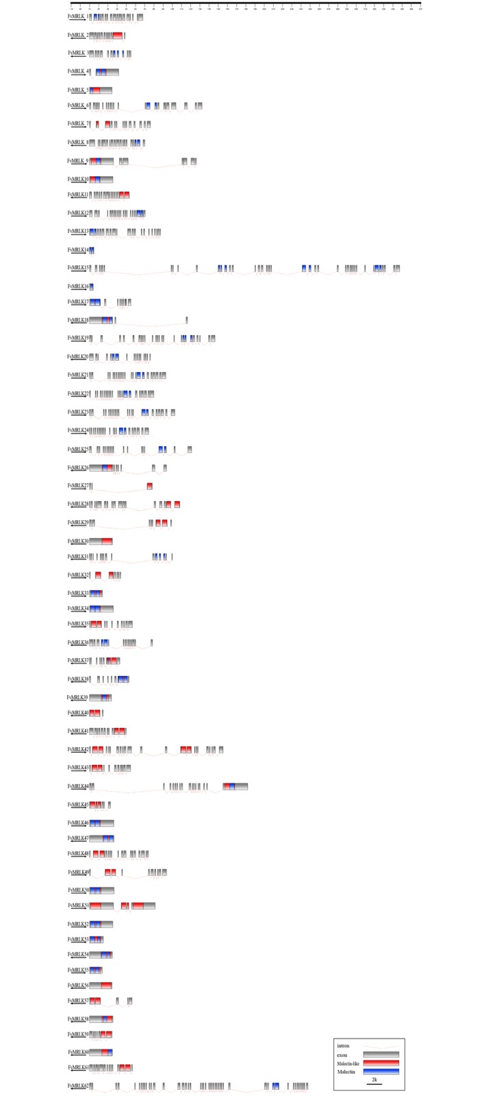
Fig 2. Structures and exon–intron composition of the FvMRLK genes.

Names of genes are indicated on the left. Exons, represented by gray boxes, are drawn to scale. Dashed lines connecting two exons represent an intron. Malectin and malectin-like domains in the FvMRLK proteins are marked in blue and red, respectively.
