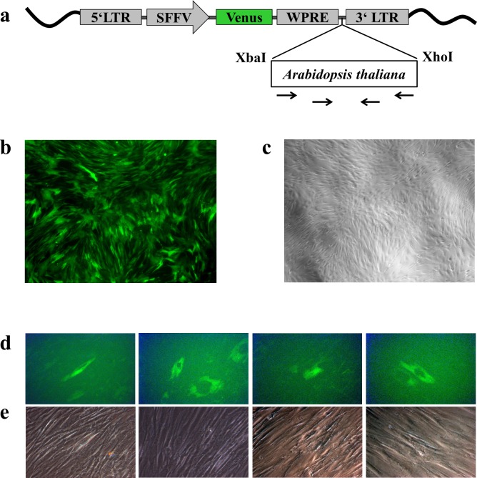
Fig 3. Arabidopsis thaliana (A.t.) virus production and transfer.
(a) A.t.-DNA was cloned into the LeGO-V2-wpre plasmid vector containing Venus-fluorescence protein for detection. Primers for subsequent primary and nested A.t.-PCR shown with arrows were located within the A.t.-sequence giving rise to products of 387 bp and 106 bp respectively. (b, c) hMSC were transduced with LeGO-V2-wpre-A.t. virus supernatant. Shown is a hMSC culture 8 days after transduction (x40) detecting green cells (b) in a near confluent culture (c, phase contrast). (d, e) Recipient hMSC were incubated for 2 weeks with EV purified from hMSC-A.t. culture supernatant. Shown are Venus-positive cells (d) in the recipient culture after incubation with EV without (3 left images) or with DNase digestion (most right image) and their respective phase contrast pictures (e) (magnification x200).

