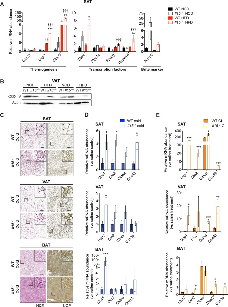
Fig 4. Il15 deficiency promotes the browning program of white adipose tissues.
(A) Quantitative PCR analysis of genes involved in thermogenesis in the SAT of WT and Il15−/− male mice fed either NCD or HFD (mean ± SEM; n = 3–4; **p<0.01, ***p<0.001 vs WT; †††p<0.001 vs NCD). (B) Immunoblot analysis for cytochrome oxidase (COX) IV and actin in VAT of 2 representative of 3 groups are shown. (C) H&E and UCP1 stained-sections of SAT, VAT and BAT of mice exposed to a cold test. Scale bar = 100 μm. Magnification ×10 (inset ×80). (D) Quantitative PCR analysis of genes involved in thermogenesis in the SAT, VAT and BAT of cold-exposed WT and Il15−/− male mice (mean ± SEM; n = 3–4; *p< 0.05). (E) Quantitative PCR analysis of genes involved in thermogenesis in the SAT, VAT and BAT of CL316243-treated WT and Il15−/− male mice (mean ± SEM; n = 3–4; *p<0.05, **p<0.01, ***p<0.001 vs WT).

