FIGURE 3:

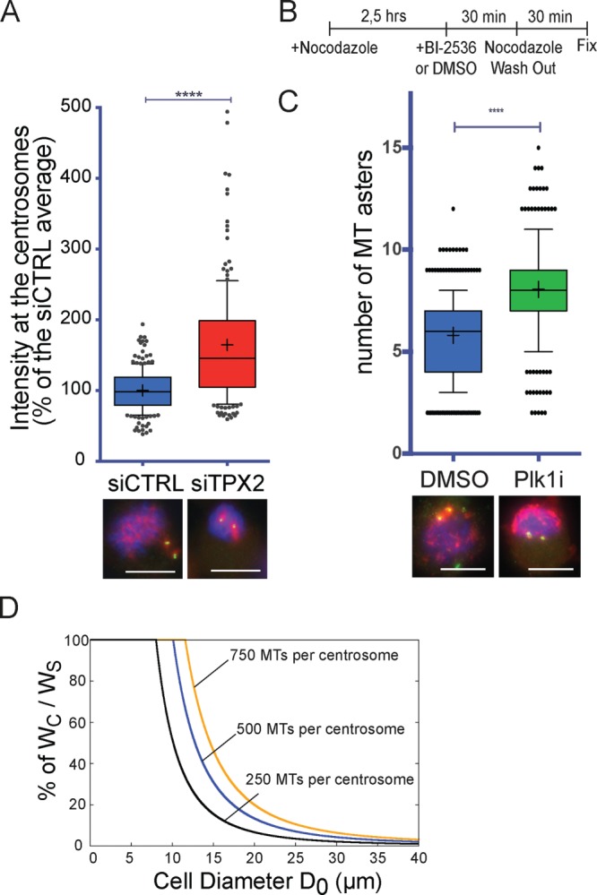
The MT assembly pathway activities are interdependent. (A) Tubulin signal on the mitotic centrosomes 1 min, 30 s after nocodazole washout in mitotic cells silenced using a scrambled siRNA (siCTRL, blue) or a TPX2 siRNA (siTPX2, red). Box-and-whiskers plot showing the intensity of the tubulin signal associated with the mitotic centrosome expressed as percentage of the average of the control. Representative IF images of siCTRL and siTPX2 are shown below the graph. DNA is in blue, centrin in green, and tubulin in red. Data from 205 siCTRL cells and 196 siTPX2 cells from four independent experiments, counting in each at least 40 cells/condition. To pool experiments, in each experiment, the mean tubulin signal of the control condition was considered as 100%. ****p < 0.0001. Scale bar, 10 μm. (B) Experimental design of the MT regrowth performed in Plk1-inhibited cells. (C) Number of MT asters generated 3 min after nocodazole washout in cells treated with DMSO (blue) or BI-2536 (Plk1i, green) as in B. Box-and-whiskers plot showing number of MT asters counted in each condition. Representative IF images of DMSO and Plk1i cells are shown below the graph. DNA is in blue, pericentrin in green, and tubulin in red. Data from 339 DMSO cells and 346 Plk1i cells from three independent experiments counting in each at least 100 cells/condition. ****p < 0.0001. Scale bar, 10 μm. (D) Percentage of the theoretical ratio of tubulin incorporated into centrosomal MTs (WC) to the total tubulin that will constitute the spindle MTs (WS), depending on cell diameter. Three theoretical levels of centrosome maturation (defined as number of MTs nucleated by the centrosomes) were modeled: 250, 500, and 750 MTs per centrosome with average MT length of 3 μm (respectively, black, blue, and orange curves). As the cell size increases, centrosomes use proportionally less of the total tubulin available for the spindle MTs. For cell diameters within 10–30 μm, altering the level of centrosome maturation has a dramatic consequence on the availability of tubulin for the chromosomal pathway. Most dividing animal cells are in this range (Milo and Phillips, 2015), and thus the model suggests that centrosome maturation at NEBD may be a tightly controlled process.
