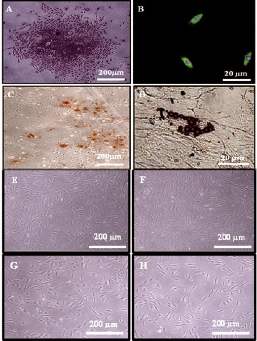
Figure 3.

(A) Phase contrast micrograph showing a colony formed by PDLSCs after staining by toluidine blue stain. (4X). (B) Phase contrast micrograph showing PDLSCs express the MSC marker STRO-1 (Green colour). Notice counter stain of the nuclei by DAPI (Blue colour). (Indirect immunocytofluorescence, 40X). (C) Phase contrast micrograph showing extracellular calcium accumulation (Orange - red nodules) by differentiated PLSCs after 14 days of osteogenic induction (Alizarin red stain, 4X), and (D) Fat droplets formed by PLSCs after 21 days of adipogenic induction (oil red O stain, 40X). Phase contrast micrograph showing PLSCs after 8 days of culturing with scaffold loaded with (E) 0% Cipro, (F) 5% Cipro, (G) 10% Cipro and (H) 20% Cipro, maintained their PLSCs morphological characteristics
