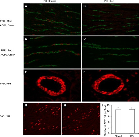
Figure 8.
Immunofluorescence validation of PRR deletion in intercalated cells of CD PRR KO mice. (A–D) The kidney sections of CD PRR KO and floxed mice were colabeled for PRR (red) and AQP2 (green) in the renal medulla. Original magnification, ×400. E and F show PRR labeling in the renal blood vessels. G and H show colabeling of AQP2 and AE1. (I) Shows the number of AE1-positive cells per field counted from 16 fields per group. All images were derived from immunofluorescence of the renal medulla.

