Figure 7.
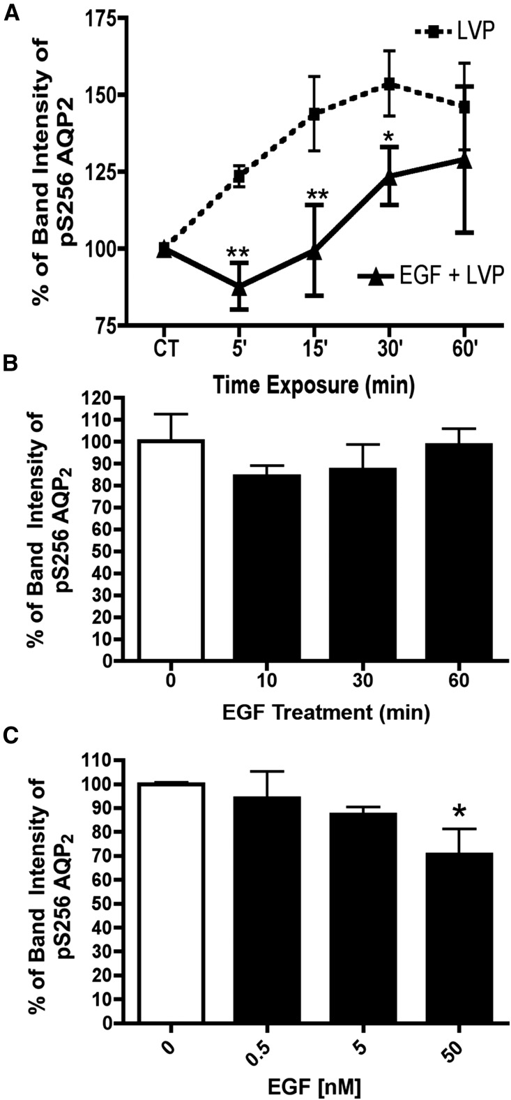
VP–induced AQP2 Ser-256 phosphorylation is inhibited by EGF. (A) LLC-PK1 cells were incubated with VP (10 nM) alone (dotted line) or coincubated with VP and EGF (5 nM; solid line) for several time periods. Phosphorylation of serine 256 was analyzed by Western blot and quantified. Data were analyzed using two–way ANOVA Bonferroni post–tests (mean±SD; n=4). EGF (5 nM) significantly decreased VP–induced Ser-256 phosphorylation of AQP2. CT, control; LVP, lysine-vasopressin. *P<0.05 compared with LVP alone; **P<0.01 compared with LVP alone. (B) EGF (5 nM) alone did not affect AQP2 S256 phosphorylation significantly after 60 minutes in LLCPK-1 cells (n=5), but (C) the effect was dose dependent. These results are the average of at least three independent experiments, and they are shown here as means±SEMs (n=3). *P<0.05 (one–way ANOVA Tukey test).

