
Figure 4.
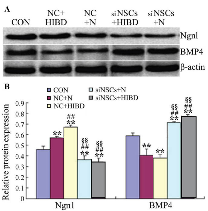
Expression levels of Ngn1 and BMP4 protein in NSCs following transfection with recombinant plasmid for 48 h. (A) Detection of Ngn1 and BMP4 protein levels by western blot analysis. β-actin was the internal standard. (B) Ngn1β-actin and BMP4β-actin protein optical density ratios were analyzed in each group. **P<0.01 vs. CON group; ##P<0.01 vs. NC+N group; §§P<0.01 vs. NC+HIBD group. Ngn1, neurogenin 1; BMP4, bone morpho-genetic protein 4; NSC, neural stem cell; CON, blank control group; NC, negative control plasmid-transfected cells; N, normal brain tissue; HIBD, hypoxic-ischemic brain damage tissue; siNSC, β-catenin small interfering RNA-transfected cell.