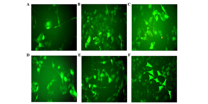
Figure 2.
Green fluorescence imaging following infection with ABMN factors. Green fluorescence imaging (A) 7, (B) 14 and (C) 21 days following infection with ABMN factors. Fraction of cells stained by green fluorescence (enhanced green fluorescence protein positive cells) (D) 7, (E) 14 and (F) 21 days following five factor (ABMN+ human brain-derived neurotrophic factor) gene transduction. All images were captured under a microscope (magnification, ×200).

