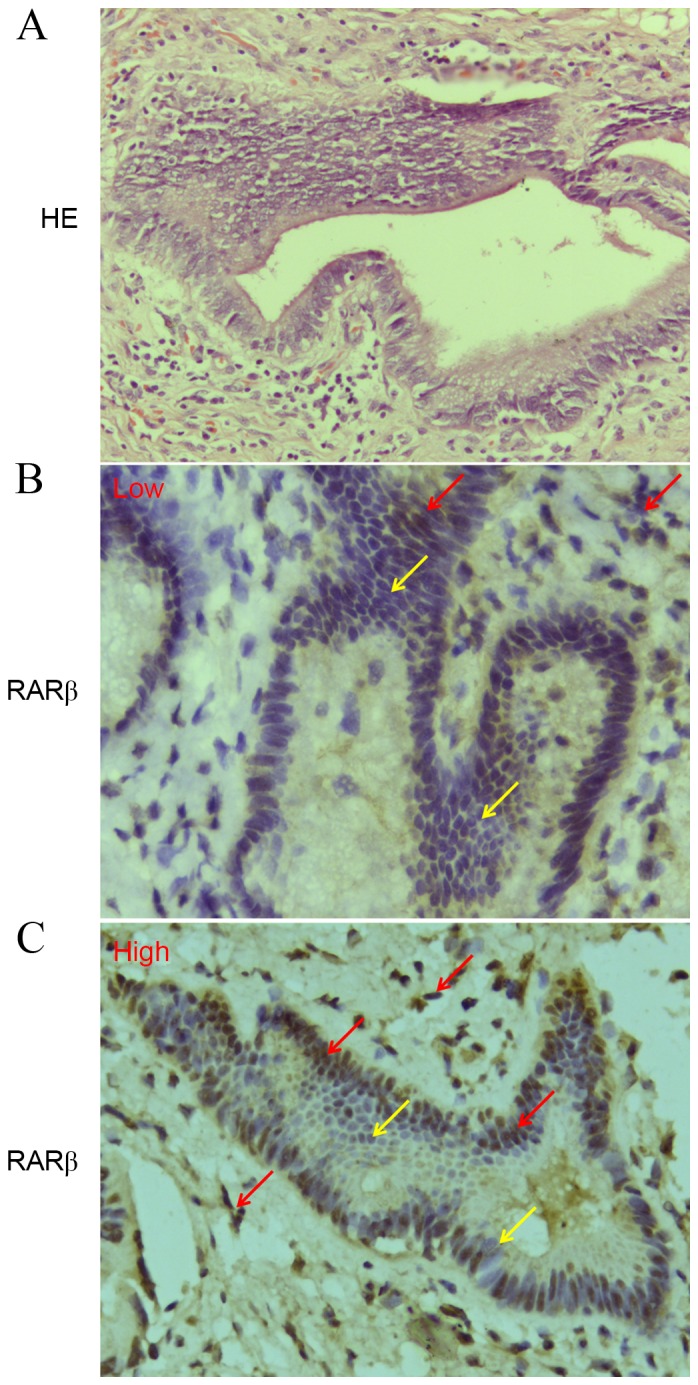
Figure 1.

Expression of RARβ protein detected by IHC in human CCA tissues. (A) HE staining. (B and C) Expression of RARβ (magnification, ×400). Yellow arrows indicate negative RARβ expression and red arrows indicate positive expression. The staining intensities of RARβ were categorized as low and high expression in 33 CCA tissues. RARβ, retinoic acid receptor-β; IHC, immunohistochemical staining; CCA, cholangiocarcinoma; HE, hematoxylin-eosin.
