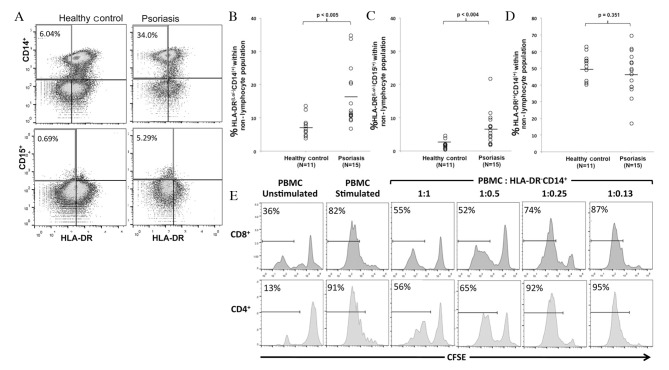
Figure 1.
MDSCs are elevated in peripheral blood from patients with psoriasis. (A) Psoriasis and healthy control PBMCs negative for the lymphocyte markers CD3, CD19, CD20 and CD56 were analyzed for HLA-DR, CD14 and CD15 expression. PBMCs isolated from patients with psoriasis had increased percentages of (B) HLA-DRlo/−CD14+ and (C) HLA-DRlo/−CD15+ cells, compared with healthy controls. (D) No significant differences were observed in the percentage of HLA-DR+CD14+ cells between the two groups. (E) CFSE-labeled PBMCs were stimulated with anti-CD3 and -CD28 in the presence or absence of autologous MDSCs at the indicated ratios. CFSE dilution indicating proliferation is presented following gating on CD4+ or CD8+ T cells. CD4+ and CD8+ T-cell proliferation was suppressed by MDSCs in a dose-dependent manner. A representative example of three experiments is presented. MDSCs, myeloid-derived suppressor cells; PBMCs, peripheral blood mononuclear cells; CD, cluster of differentiation; HLA, human leukocyte antigen; CFSE, 5-(and 6) carboxyfluorescein diace-tate succinimidyl ester.

