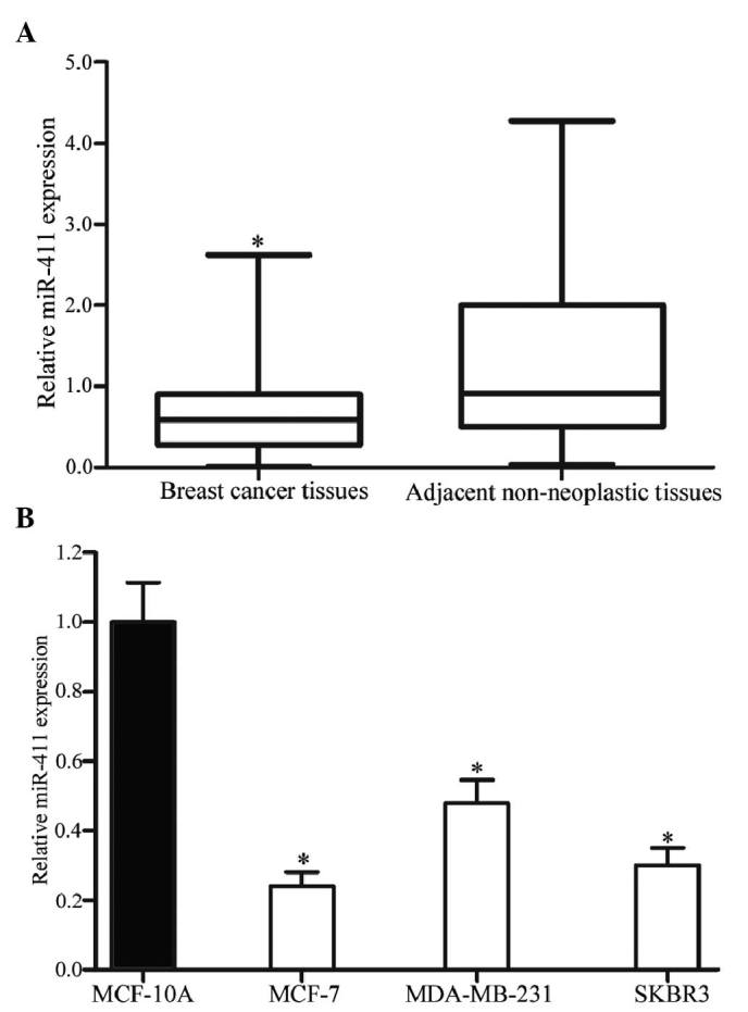
Figure 1.

Expression levels of miR-411 in breast cancer tissues and cell lines. (A) Levels of miR-411 in the breast cancer tissues were significantly lower than in their corresponding adjacent non-neoplastic tissues. (B) Levels of miR-411 were also lower in the MCF-7, MDA-MB-231 and SKBR3 cell lines, compared with the MCF-10A cell line. Expression levels of miR-411 were determined by reverse transcription-quantitative polymerase chain reaction analysis and normalized to U6. Data are presented as the mean ± standard deviation. *P<0.05, compared with their respective controls. miR, microRNA.
