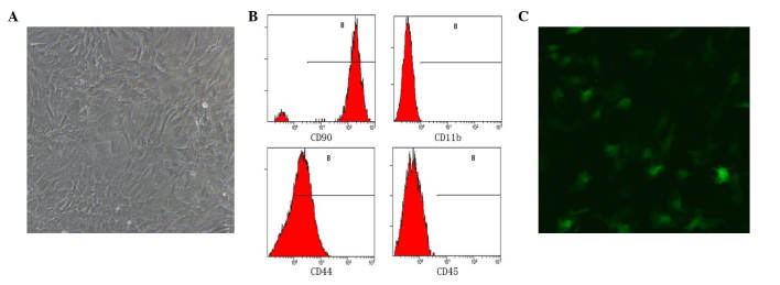
Figure 1.
Bone marrow-derived mesenchymal stem cells under fluorescence microscopy (magnification, ×20). (A) Cells are attached to the bottom of the culture dish exhibit a spindle shape. (B) Flow cytometry results, cells express CD90 and CD44 and are negative for CD45 and CD11b. (C) Green fluorescent protein positive cells are observable under fluorescence microscopy. CD, cluster of differentiation.

