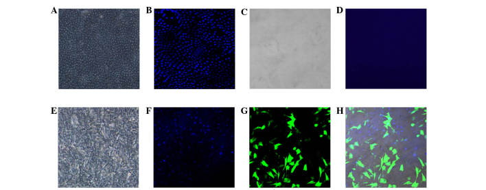
Figure 2.
Amniotic membrane with cultured stem cells under confocal microscope (magnification, ×40). (A) Normal amniotic membrane with epithelial layer, (B) epithelial cell nuclei stained with DAPI demonstrated regular arrangement; (C) denuded amniotic membrane with (D) no visible DAPI stained nuclei; (E) amniotic membrane with cultured stem cells; (F) nuclei of stem cells stained with DAPI demonstrated a irregular arrangement; (G) stem cells expressing GFP protein; (H) GFP-positive cells with DAPI stained nuclei. DAPI, 4′,6-diamidino-2-phenylindole; GFP, green fluorescent protein.

