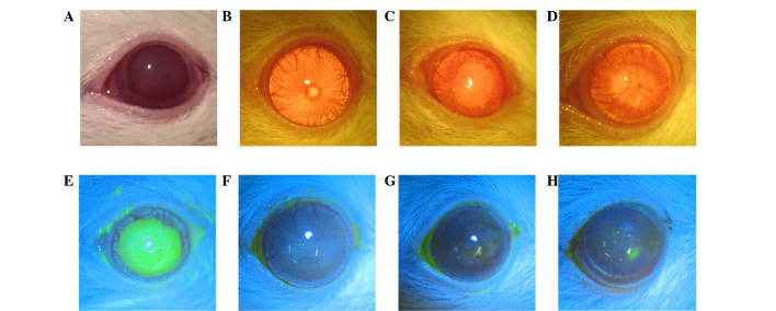
Figure 3.
Corneal surface evaluation prior to and 4 weeks after treatments. (A–H) Images of rat corneas. (A) Cornea on the day of injury, (E) total epithelial defect stained with fluorescein dye; (B and F) Group 1, clear, smooth cornea without epithelial defects or vessels following 4 weeks of subconjunctival injection; (C and G) Group 2, cornea with irregular surface and new vessels 4 weeks following amniotic membrane transplantation; (D and H) Group 3 (control), small epithelial defect stained with fluorescein dye and new vessels visible.

