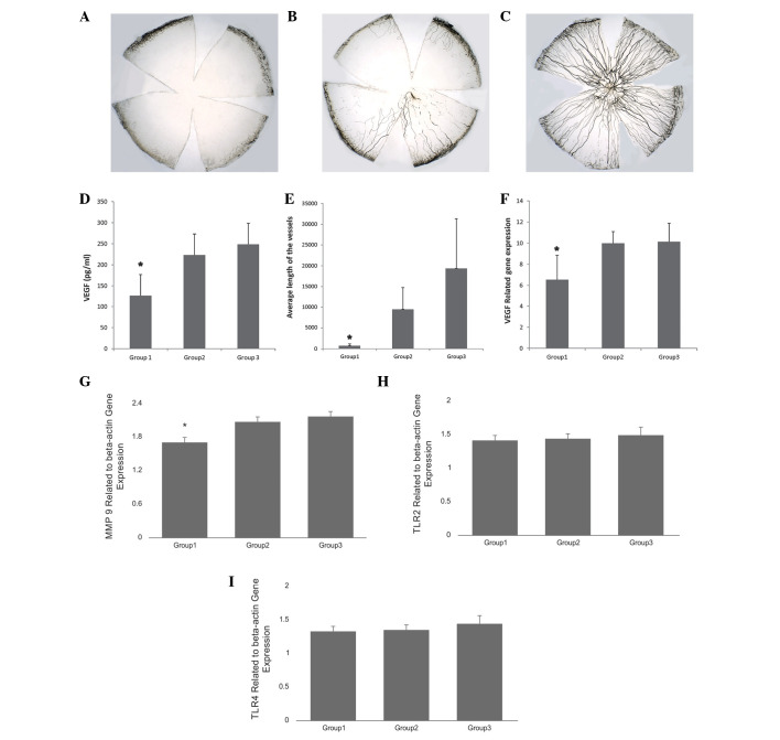
Figure 4.
Corneal flat mounts under light microscope (magnification, ×10). (A) Subconjunctival injection group (group 1), vessels are short and only in one quadrant; (B) amniotic membrane transplantation group (group 2), vessels are in more than half of the cornea and reach to the center; (C) control group (group 3), vessels are over the entire cornea; (D) VEGF protein level detected by ELISA; (E) length of the vessels measured by Image-Pro Plus 6.0; (F) VEGF, (G) MMP-9, (H) TLR2 and (I) TLR4 gene expression relative to actin. *P≤0.05, 95% confidence interval. VEGF, vascular endothelial growth factor.

