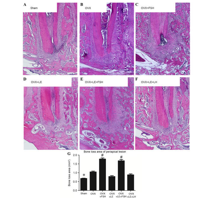
Figure 2.
Histological analysis of periapical lesions in each group. (A) Sham, (B) OVX, (C) OVX + FSH, (D) OVX + LE, (E) OVX + LE + FSH and (F) OVX + LE + LH groups. (G) Quantitative analysis of periapical lesions in each group. Compared with those in the Sham group, specimens in the OVX groups exhibited significant increases in bone loss of periapical lesions (P<0.05; A, B and G), which were reversed by the administration of LE or LE + LH (P>0.05; D–G). Bone loss of periapical lesions was significantly increased vs. those in the Sham and OVX groups after administration of FSH (P<0.05; C, E and G). Data is presented as the mean ± standard deviation. *P<0.05 vs. the OVX only group and the FSH treatment group; #P<0.05 vs. non-FSH treatment groups. OVX, ovariectomised; FSH, follicle-stimulating hormone; LE, leuprorelin; LH, luteinizing hormone.

