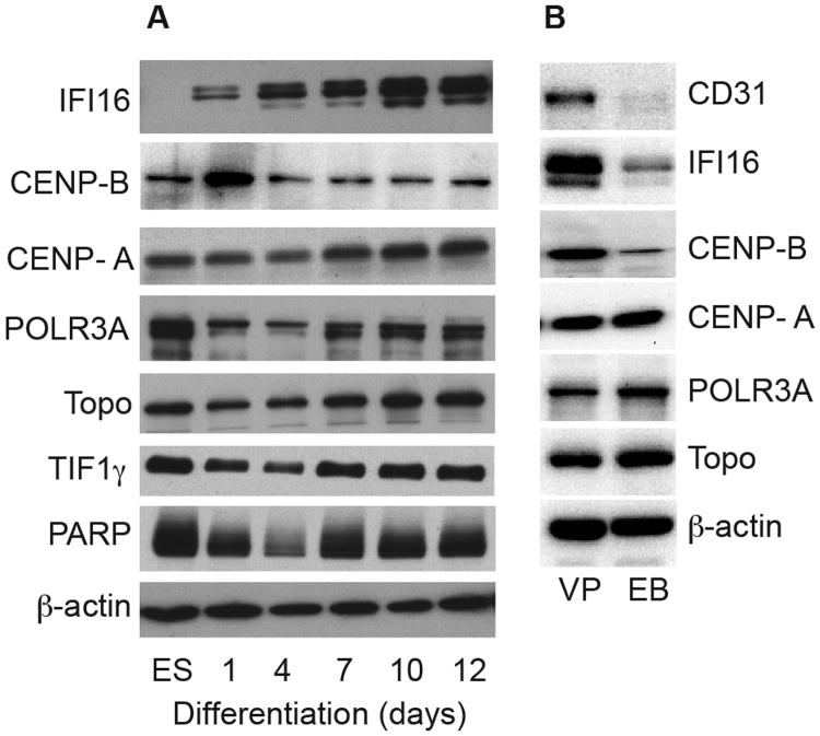
Figure 2. Scleroderma autoantigen expression levels in the progenitor cell differentiation system.
(A) Lysates made from human ES cells and EBs harvested at different times of differentiation over a 12-day period were immunoblotted with commercial antibodies against IFI16, CENP-B, POLR3A, topoisomerase I, and TIF1γ, or patient sera with high titer antibodies against PARP or CENP-A. (B) Lysates made from day 12 EBs and vascular progenitors (“VP”) were immunoblotted as above; a rabbit polyclonal antibody was used for the CENP-A blot. (A & B): Equal protein amounts were electrophoresed in each gel lane. The same lysates were immunoblotted with an antibody against β–actin as a loading control (lowest panels of each set).

