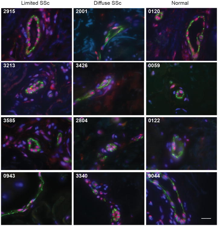
Figure 4. IFI16 expression is enriched in vascular endothelial cells in skin biopsies from both normal individuals and scleroderma patients.
Skin paraffin sections from 20 scleroderma patients (9 with limited disease and 11 with diffuse disease) and 8 normal controls were double stained with a rabbit polyclonal antibody against CD31 (depicted in green) and a mouse monoclonal anti-IFI16 antibody (depicted in red). All sections were counterstained with DAPI (represented in blue). Representative data (merged images) from 4 scleroderma patients with limited disease (tissue numbers 2915, 3213, 3585 and 0943), 4 scleroderma patients with diffuse disease (tissue numbers 2001, 3426, 2804, and 3340) and 4 normal controls (tissue numbers 0120, 0059, 0122 and 9044) is shown. Robust levels of IFI16 staining are found in vascular endothelial cells in both normal controls and scleroderma patients. Scale bar represents 20 microns. Tissues 2915, 3213, 2001 and 3426 are from patients with digital ulcers or digital gangrene; all other scleroderma tissue was from patients with Raynaud's phenomoneon without digital ulcers.

