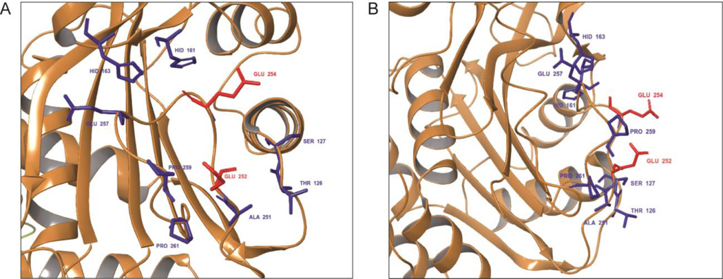
Figure 2.
Constellation of surface protruding residues located in the binding region comprising residues 246–264 of the lower pH conformation of cdb3. Accessible surface amino acids residing within 12Å of the geometric center of the above sequence were determined from the crystal structure of cdb3, and are shown with an (A) en face view and (B) right-rotated view. The two most exposed residues, Glu 252 and Glu 254, are highlighted in red. Less prominently exposed residues are highlighted in blue.

