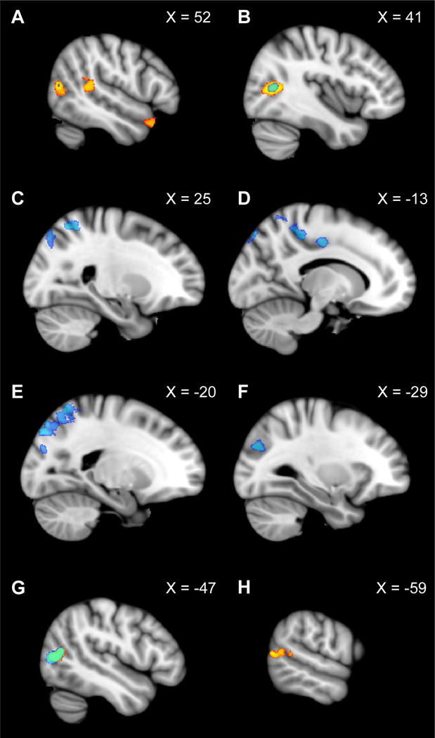
Figure 4.

Group analysis exploring representation first-person perspective motion information beyond OPA. The contrast of “Dynamic Scenes > Static Scenes” is shown in blue (p < 0.01, FWE corrected), while the contrast of “Dynamic Faces > Static Faces” is shown in yellow (p < 0.01, FWE corrected). The right hemisphere is depicted in panels A–C, while the left hemisphere is depicted in panels D–H. X coordinates in MNI space are provided for each slice. A network of regions including lateral superior occipital cortex (corresponding to OPA; see F), superior parietal lobe, and precentral gyrus (see C–E) responded significantly more to “Dynamic Scenes > Static Scenes” (blue), but similarly to “Dynamic Faces vs. Static Faces” (yellow). One bilateral region in lateral occipital cortex (corresponding to motion-selective MT) showed overlapping activation across both contrasts (see B, G). Finally, regions in bilateral posterior superior temporal sulcus and anterior temporal pole responded more to “Dynamic Faces > Static Faces,” but similarly to “Dynamic Scenes vs. Static Scenes”, consistent with Pitcher et al. (2011) (see A and H).
