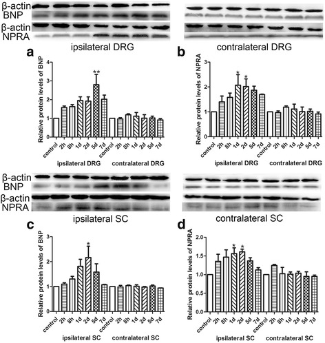
Fig. 2.

Western blot analysis showed the protein expression of BNP and NPRA in DRG and spinal cord at different time points after BmK I injection. a: The protein expression of BNP in bilateral sides of L4-L5 DRG. b: The protein expression of NPRA in bilateral sides of L4-L5 DRG. c: The protein expression of BNP in bilateral sides of L4-L5 spinal cord. d: The protein expression of NPRA in bilateral sides of L4-L5 spinal cord. *P < 0.05, **P < 0.01, ***P < 0.001 compared with control groups (n = 3). Error bars indicated S.E.M
