Fig. 5.

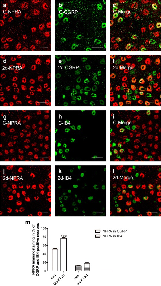
Representative microphotographs showed the location and expression of NPRA in CGRP-positive and IB4-positive small DRG neurons. Immunofluorescene staining for NPRA (a, d) and CGRP (b, e) were co-localized (c, f) in control (a-c) at 2 days after i.pl. BmK I injection (d-f). Immunofluorescene staining for NPRA (g, j) and IB4 (h, k) were co-localized (i, l) in control (g-i) at 2 days after i.pl. BmK I injection (j-i). m, Ratio of CGRP-positive and IB4-positive small DRG neurons co-localized with NPRA between control group and the group of 2 days after i.pl. BmK I injection. Scale bar, 50 μm
