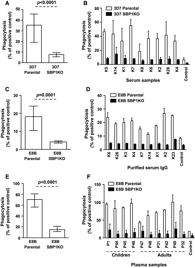
Fig. 6.
Opsonic phagocytosis of IEs by undifferentiated Thp-1 monocytes. Opsonic phagocytosis activity of serum antibodies from Kenya and PNG was markedly reduced with 3D7 SBP1KO (a) and E8B SBP1KO (c, e) compared to 3D7 parental and E8B parental. Assays were performed twice independently; bars represent median and interquartile ranges [for Kenyan samples, n = 25 for 3D7 (a), n = 9 for E8B (b); for PNG samples, n = 25 for E8B (c)]; p value was calculated using a paired Wilcoxon signed rank test. For all graphs, the level of opsonic phagocytosis is expressed as a percentage of positive control (rabbit antibody raised against human erythrocytes). A representative selection of serum samples from Kenya tested for opsonic phagocytosis activity to 3D7 SBP1KO (b) and E8B SBP1KO (d) and from PNG tested for opsonic phagocytosis activity to E8B SBP1KO (f). Assays were performed twice independently for 3D7 and once for E8B; bars represent mean and range with samples tested in duplicate

