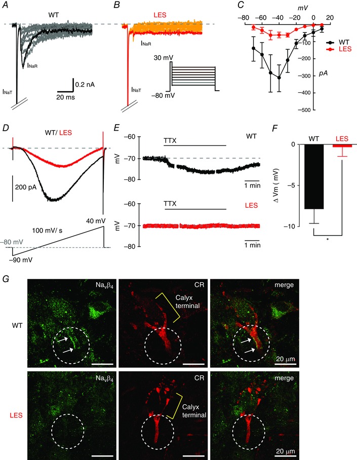
Figure 5. Reduced resurgent and persistent Na+ currents at axon terminal in the LES rat .

A and B, representative traces of resurgent Na+ currents evoked by step repolarization (from +20 mV to −70 mV) after a brief depolarization (+30 mV, 5 ms) to generate transient Na+ currents (INaT) at the calyx of Held terminals in WT (A) and LES rats (B). C, current–voltage (I–V) relationship of resurgent Na+ currents form WT (black) and LES rats (red). D, persistent Na+ currents were recorded by ramp depolarization from −90 mV to +40 mV (100 mV/s) at the calyx of Held terminals in WT (black) and LES rats (red). Representative traces of persistent Na+ currents after TTX subtraction. E, in current clamp recordings, TTX (1 μm) hyperpolarized the calyx terminal in the MNTB from WT but not LES rats. F, summary of changes in calyx membrane resting potential (ΔV m) by the application of TTX (1 μm) in the MNTB from WT and LES rats. G, Navβ4 (green) expression at the axon heminode with calretinin (CR, red) in WT and LES rats. Circles indicate the heminodal region and arrows indicate the Navβ4 cluster. Unpaired t test, * P < 0.05. [Colour figure can be viewed at wileyonlinelibrary.com]
