
Figure 3. Examples of effects of auxiliary subunits on CaV2.2 calcium channel currents .
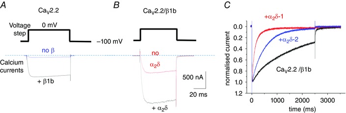
A, CaV2.2 calcium currents: effect of β subunits. Example of peak CaV2.2 current at 0 mV in the absence of β (blue) and presence of β1b (black). B, CaV2.2 calcium currents: effect of α2δ subunits. Example of peak CaV2.2/β1b current at 0 mV in the absence of α2δ (red) and presence of α2δ‐3 (black). Scale bars apply to both A and B. Charge carrier 1 mm Ba2+, expression in tsA‐201 cells, as in a previous study (Leroy et al. 2005). C, effect of different α2δ subunits on inactivation. Examples of normalized peak current for CaV2.2–β1b (black), CaV2.2–β1b–α2δ‐2 (blue) and CaV2.2–β1b–α2δ‐1 (red), over a 2.5 s timescale. Expression in Xenopus oocytes, as in a previous study (Canti et al. 2005).