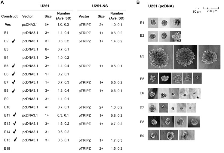
Figure 2. Soft agar colony formation assay.
A. colony number of U251 stable transfectants and U251-NS lentiviral infectants of wild-type EFEMP1 and EFEMP1 variants, normalized to the average number of colonies from the Vec control (set at unity). The average (Ave) and standard deviation (SD) were based on colonies formed in 4 individual wells. Representative colonies (5-10), were measured and scaled by the diameter of the colony: 1+ (~10 μm), 2+ (~20 μm), 3+ (30-40 μm), 4+ (40-60 μm), 5+ (50-100 μm), 6+ (100-200 μm). B. representative images of colonies from U251 stable transfectants.

