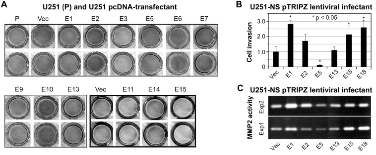
Figure 3. Detection of cell invasiveness.
A. a photograph of bottom layers in transwells from a matrigel cell invasion assay, with duplicates of cells of U251 stable transfectants of wild-type EFEMP1 and EFEMP1 variants. Invasion pictures from two independent experiments were shown by think and thick-line boxes. B. a plot of normalized cell counts in bottom layers of transwells from a matrigel cell invasion assay, with triplicates of cells of U251-NS lentiviral infectants of wild-type EFEMP1 and EFEMP1 variants, following a 3-day culture in doxycycline-containing medium. C. a photograph of two gels of the zymography assay, conducted with cells from the matrigel invasion assay described in panel B.

