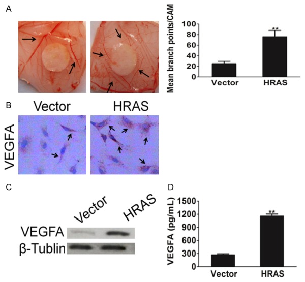
Figure 3.

HRAS facilities tumor cells induced angiogenesis. A. Representative Images of CAM blood vessels stimulated with conditioned medium from MKN28 cells. B. A representative cell immunohistochemistry assay shown VEGFA protein expression in control cells and MKN28 cells overexpression HRAS. C. Western blot shown that VEGFA was elevated in cells transfected with vector or HRAS retrovirus plasmid. β-Tublin was used as a loading control. D. MKN28 cells grown to 70-90% confluence were co-transfected with vector or HRAS plasmid. Cell culture supernatants from indicated cells were performed by ELISA assay for assay VEGFA mount. Each bar represents the mean ± SD of three independent experiments. *P < 0.05, **P < 0.01.
