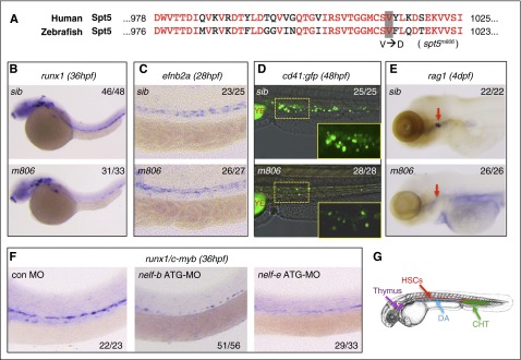
Figure 1.
Defective HSC formation in pausing-deficient embryos. (A) Alignment of the C-terminal protein sequence of human and zebrafish Spt5. The V1012D mutation in the zebrafish spt5m806 mutant is highlighted. (B-C) WISH in sibling control (sib) and spt5m806 (m806) embryos for expression of runx1 at 36 hpf (B) and ephrinb2a (efnb2a) at 28 hpf (C). (D) Overlay of the bright field and fluorescent imaging of CHT in Tg(cd41:GFP) embryos at 48 hpf. The insets depict the enlarged GFP fluorescent imaging of the area outlined by dashed lines. YE, yolk extension. (E) WISH for rag1 in thymus (red arrows) at 4 dpf. (F) WISH for runx1/c-myb at 36 hpf in embryos injected with control morpholino (MO), nelf-b MO, or nelf-e MO. (G) Illustration of the blood organs analyzed in panels B-F. Numbers in the upper/lower right corner of images indicate the fraction of embryos with WISH signal similar to the representative image.

