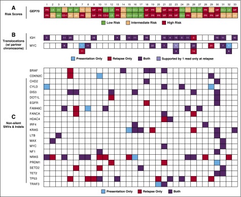
Figure 1.
GEP70 risk, IgH, and MYC translocations and nonsilent mutations in selected genes. (A) The risk status according to the recently published 3-group GEP70 classifier11 is shown for the 33 patients. Intermediate-risk cases would be classified as low risk according to the classical GEP70.10 UAMS GEP groups are shown within the boxes, consisting of CD-1, CD-2, HY, LB, MS, MF, and PR. The upper and lower rows present the status at presentation and relapse, respectively. (B) IgH and MYC translocations were called from whole-exome sequencing data using MANTA16 and are shown with the corresponding translocation partner chromosome. C: Non-silent SNVs and indels in a set of genes previously implicated in the pathogenesis of MM.17,18 CoLRs indicate whether an abnormality was detected at presentation, relapse, or both.

