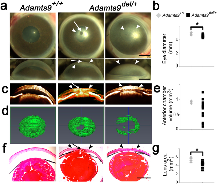
Figure 1. Adamts9del/+ mice have a highly penetrant congenital corneal opacity resulting from ASD.
(a–g) Corneal opacity in Adamts9del/+ eyes is associated with Peters anomaly (a), a smaller eye (b), shallow anterior chamber (c–e), iridocorneal and iridolenticular adhesions (f) and a smaller lens (g). 3 week-old Adamts9+/+ and Adamts9del/+ enucleated eyes were analyzed by stereomicroscopy (a), OCT (c,d) and H&E staining of paraffin sections (f). The arrowheads and arrow indicate, respectively, the corneal opacity and the Peters anomaly. Adamts9del/+ eyes had a significantly smaller diameter than Adamts9+/+ eyes (b). Anterior chamber volume was determined from OCT data using the Amira segmentation tool (d). Anterior chamber volumes were highly variable in Adamts9del/+ eyes while quite constant in Adamts9+/+ eyes (e). Lens area was measured from H&E stained sections and was significantly smaller in Adamts9del/+ eyes as compared to Adamts9+/+ eyes (g). The images are representative of 6 Adamts9+/+ and 12 Adamts9del/+ eyes analyzed by OCT. Scale bar = 1 mm. Significance was determined using a 2-tailed student’s t test (*p < 0.05).

