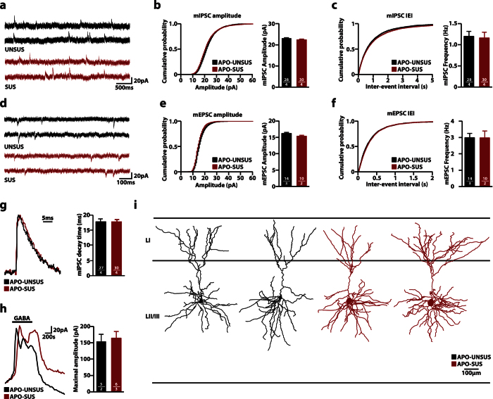
Figure 3. No changes in basal synaptic transmission and morphology in LII/III pyramidal cells in the medial prefrontal cortex (mPFC).
(a) Representative traces of miniature inhibitory post-synaptic currents (mIPSCs) recorded from LII/III pyramidal cells from acute brain slices from APO-UNSUS and APO-SUS rats. (b,c) Cumulative distributions of mIPSC amplitudes (b) and inter-event intervals (IEI) (c) from APO-UNSUS and APO-SUS rats. (d) Representative traces of miniature excitatory post-synaptic currents (mEPSCs) recorded from LII/III pyramidal cells from acute brain slices from APO-UNSUS and APO-SUS rats (e,f) Cumulative distributions of mEPSC amplitudes (e) and inter-event intervals (f) from APO-UNSUS and APO-SUS rats. (g) Representative traces (scaled) and distribution of mIPSC decay time. (h) Representative traces and quantification of systemic GABA (20 μM) application from APO-UNSUS and APO-SUS rats. Black bar indicates GABA application. (i) Representative reconstructions of LII/III pyramidal neurons from mPFC. Sample size (n) is indicated in the bars as number of cells/number of animals. Bar graphs are presented as mean ± SEM. Student’s t-test.

