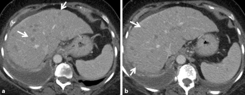
Fig. 1.
Case 2: a 65-year-old woman with ER/PR(+) infiltrating ductal adenocarcinoma of the right breast. Contrast-enhanced axial CT images 2 years following mastectomy and adjuvant chemoradiation (a, b) show an enlarged fatty liver with several small hypodense round masses in both the right and left hepatic lobes corresponding to diffuse metastatic disease (white arrows in a, b).

