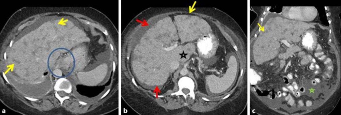
Fig. 2.
Case 2: follow-up contrast-enhanced CT in the same patient 6 months after initiation of paclitaxel and gemcitabine, followed by vinorelbine, ixabepilone and capecitabine showed features of pseudocirrhosis. Contrast-enhanced axial (a, b) and coronal (c) images show several ill-defined, somewhat linear, ‘band-like’ masses that extend to the periphery of the liver (yellow arrows in a–c). There is secondary capsular retraction and nodularity of the liver surface (red arrows in a, b) and an enlarged caudate lobe (black star in b). Changes of portal hypertension are present with paraesophageal varices (blue circle in a) and new ascites (green star in c).

