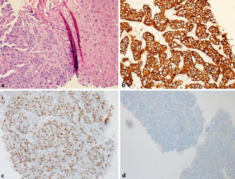
Fig. 4.
a Fine needle aspiration biopsy of the liver normal parenchyma of the liver (right) with tumor infiltration with malignant cells (left). ×200. b, c Immunohistochemical staining of the liver tumor shows positive staining for Her2 3+ (×200) and positive staining for GATA-3 (×200). d Negative control for Her2 and GATA. ×100.

