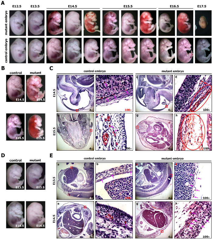
Figure 2. The EIIa-Cre-mediated activation of Cripto-1 leads to hemorrhaging and embryonic lethality.
(A) Whole-mount views of representative control (EIIa-Cre) and mutant (RCLG/EIIa-Cre) embryos at E12.5 to E17.5. (B,C) Histological analysis of control (a,b,e,f) and mutant (c,d,g,h) embryos at E14.5 and E15.5. E14.5 and E15.5 mutant embryos (B) demonstrated hemorrhages. Sagittal sections of E14.5 control and mutant embryos (indicated in B) were stained with H&E (a–d), while transverse sections of E15.5 control and mutant embryos (indicated in B) were stained with H&E (e–h). In Fig. 2C(b,d,f,h) are higher magnifications of the red rectangular regions indicated in (a,c,e,g), respectively. The pink arrows indicate normal capillaries (b,f) with erythrocytes in the control embryos. The red ovals in (d,h) outline hemorrhaging of the body surface capillaries of mutant embryos, while the red arrow shows a normal great vessel (d) in a mutant embryo. (D,E) Histological analysis of control (a,b,e,f) and mutant (c,d,g,h) embryos at E13.5 and E14.5. No significant difference was found in the body surfaces of E13.5 control and mutant embryos (D, upper image), whereas the E14.5 mutant embryo appears pale compared to the E14.5 control embryo (D, lower image). Sagittal section of E13.5 and E14.5 control and mutant embryos (indicated in D) were stained with H&E. In Fig. 2E,(b,d,f,h) are higher magnifications of the red boxes indicated in (a,c,e,g), respectively. The pink arrows indicate normal capillaries (b,d,f,h) with or without erythrocytes, which are nucleated in both control and mutant embryos at this stage of development.

