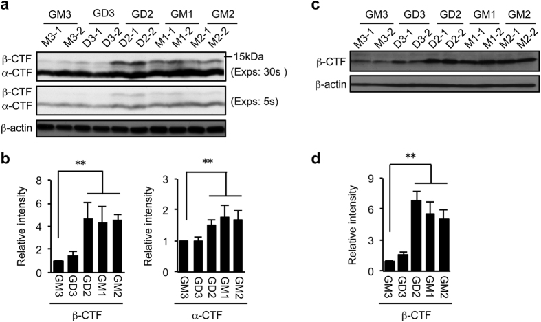
Figure 2. Effect of ganglioside expression on APP processing.
Whole cell lysate was prepared from cells treated with 1 μM DAPT for 12 h. Equal amounts of cellular proteins were subjected to immunoblot analysis with anti-APP antibody Y188 (a) or 82E1(c). Longer (30 sec) and shorter (5 sec) exposure images are shown in panel a. β-actin served as an internal standard. Levels of α-CTF and/or β-CTF shown in panels a and c were quantified and presented in panels b and d, respectively. Data represent means ± s.d. (n = 6). Statistical analysis was performed by one-way ANOVA with Tukey-Kramer post hoc test (**P < 0.01).

