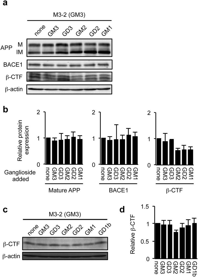
Figure 4. Effect of exogenous gangliosides on BACE1 expression and activity.
(a,b) BACE1 protein expression. GM3-expressing cells (M3-2) were incubated without or with GM3, GD3, GM2, GD2 or GM1 (30 μM each) for 21 h. Whole cell lysate was subjected to immunoblot analysis for BACE1, APP (22C11) and β-CTFs (Y188). β-actin served as an internal standard. BACE1, APP and β-CTF levels were quantified and plotted in panel b. Data are means ± s.d. (n = 3). Statistical analysis was performed by one-way ANOVA with Dunnett post hoc test, and statistical significance was not detected. IM, immature form; M, mature form. (c,d) Generation of β-CTF. GM3-expressing cells (M3-2) were treated with gangliosides as above. Twelve hours before harvesting cells, DAPT was added to culture medium at the final concentration of 1 μM. Whole cell lysate was subjected to immunoblot analysis with anti-Aβ (82E1) that detects only β-CTF. β-CTF level was quantified and plotted in panel d. Data represent means ± s.d. (n = 3). Statistical analysis was performed by one-way ANOVA with Dunnett post hoc test, and statistical significance was not detected.

