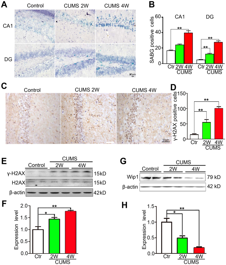
Figure 4. CUMS experience increased cellular senescence and γ-H2AX activity, but reduced Wip1 expression in wildtype mice.
(A) Illustration of SABG-staining positive cells within CA1 and DG subareas of those wildtype mice exposed to CUMS for 2 or 4 weeks, or left undisturbed (the control group). (B) CUMS experience significantly increased cellular senescence within CA1 and DG subareas in a time-dependent manner (n = 6 mice for each group). (C) Illustration of hippocampal γ-H2AX positive cells in CUMS-exposed mice and their controls. (D) CUMS experience significantly increased the number of γ-H2AX positive cells in hippocampus compared to their controls (n = 6 mice for each group). (E) The expressions of γ-H2AX and the total H2AX in hippocampal tissues of CUMS-exposed mice and their controls were detected using Western blotting assay. (F) CUMS experience significantly increased the expression of γ-H2AX in hippocampus compared to their controls (n = 6 samples for each group). The expression of γ-H2AX was normalized to that of the total H2AX. (G) The expression of Wip1 in hippocampal tissues of CUMS-exposed mice and their controls was detected using Western blotting assay. (H) CUMS experience significantly reduced the Wip1 expression in hippocampus when compared to their controls (n = 6 samples for each group). Wip1 was normalized to β-actin. *P < 0.05; **P < 0.01.

