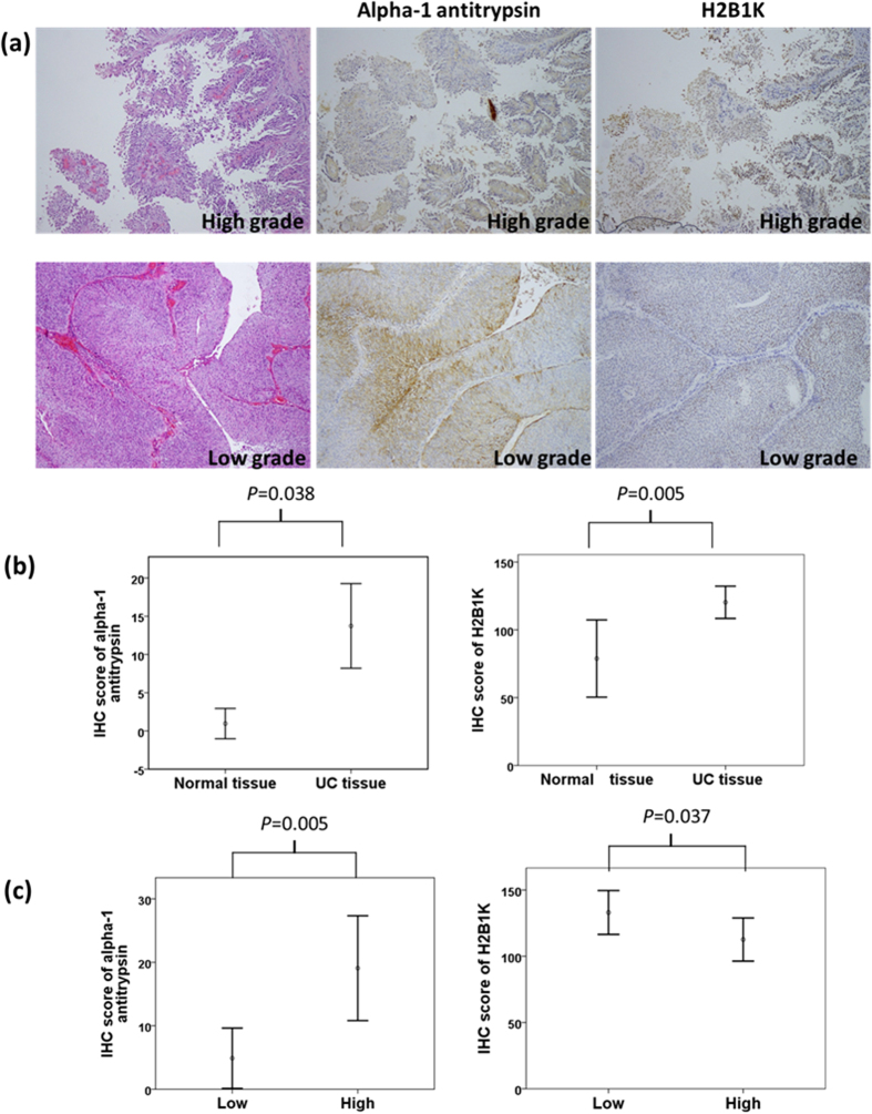
Figure 5.
(a) Hematoxylin and eosin staining of high-grade (upper panel) and low-grade (low panel) UC tissues with the corresponding IHC staining of alpha 1-antitrypsin and H2B1K. Alpha 1-antitrypsin and H2B1K were subjected to cytoplasmic and nuclear staining, respectively. (b) Overexpression of alpha 1-antitrypsin and H2B1K in UC tissues was compared with that in normal tissues. (c) Different expression levels of alpha 1-antitrypsin and H2B1K in high-grade UC tissues were compared with those in low-grade UC tissues.

