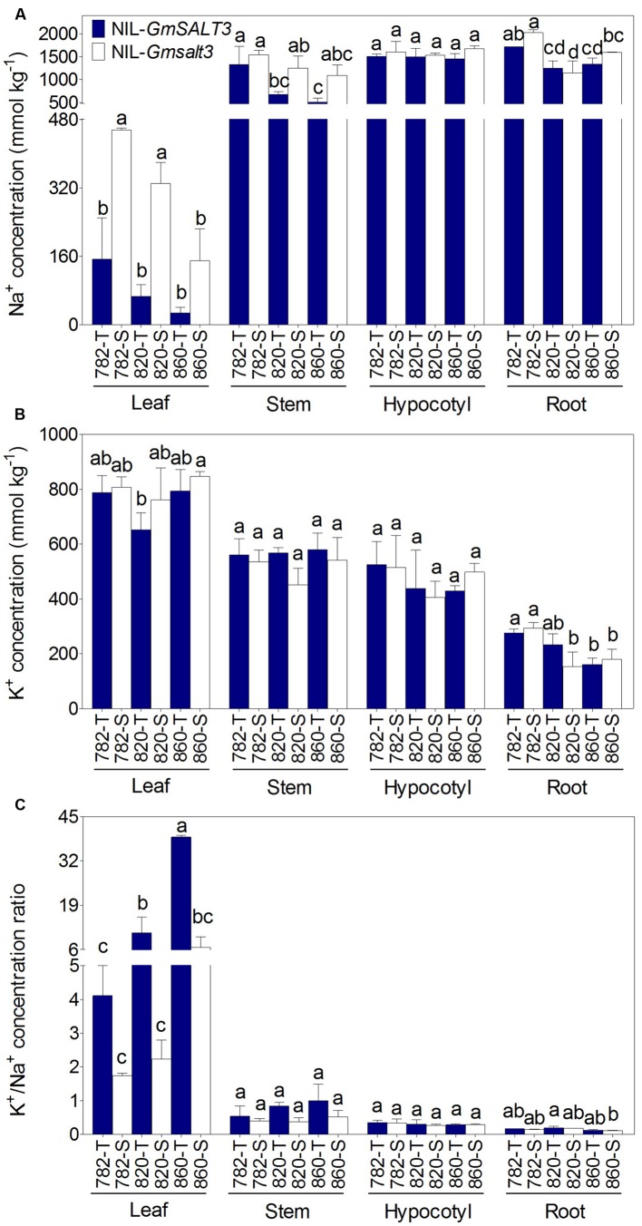
FIGURE 3.
Ion concentration (dry mass) in three sets of NILs after 10 days of NaCl stress (200 mmol L-1; EC = 17.8 dS m-1). (A) Tissue concentration of Na+ in leaf, stem, hypocotyl and root of three sets of NILs. (B) Concentration of K+ in leaf, stem, hypocotyl and root of three sets of NILs. (C) The K+/Na+ ratio in leaf, stem, hypocotyl and root of three sets of NILs. Data are means of three replicates consisting of the mean values for five plants grown in the same pot ± SD (n = 3). Different letters indicate statistically significant differences between NIL lines for each tissue (one-way ANOVA followed by Tukey’s HSD post hoc test, P < 0.05).

