Abstract
The conventional technique of impression cytology provides a non-invasive method for the evaluation of conjunctival epithelium alterations. Using indirect immunofluorescence procedures two inflammatory markers, class II MHC antigens HLA DR and the receptor to IgE (CD23), were sought in impression cytology specimens obtained from 80 patients. In normal subjects conjunctival epithelial cells did not show any reactivity. Only scattered dendritic cells were found to express class II antigens but not the receptor to IgE. In contrast patients with chronic conjunctivitis of various aetiologies, mainly infectious or allergic, had 40-100% of brightly positive conjunctival cells for one or both antigens. In these cases epithelial cells and goblet cells reacted similarly. Twenty four eyes in 12 patients with idiopathic dry eye syndrome disclosed results similar to those from normal conjunctival specimens. However 18 other specimens from patients suffering from idiopathic tear deficiency but undergoing multiple substitutive treatments for dry eye had moderate to strong positivity for HLA DR and/or the receptor to IgE (20-100% of cells). As these results were independent of the degree of squamous metaplasia the expression of these membrane markers may reflect local inflammation in addition to tear deficiency, possibly due to sensitisation to the eye drops used. These immunocytological techniques thus provide useful methods of investigating conjunctival inflammation and allergy. They may constitute valuable aid in the diagnosis and appropriate treatment of ocular surface disorders.
Full text
PDF
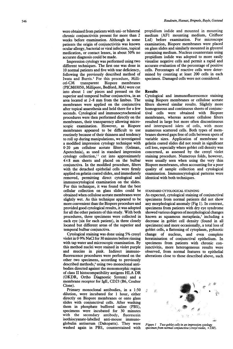
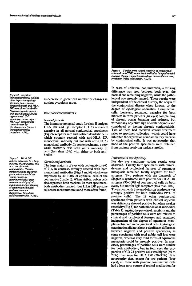
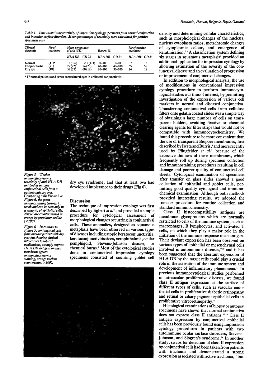
Images in this article
Selected References
These references are in PubMed. This may not be the complete list of references from this article.
- Baudouin C., Fredj-Reygrobellet D., Gastaud P., Lapalus P. HLA DR and DQ distribution in normal human ocular structures. Curr Eye Res. 1988 Sep;7(9):903–911. doi: 10.3109/02713688808997247. [DOI] [PubMed] [Google Scholar]
- Baudouin C., Fredj-Reygrobellet D., Gordon W. C., Baudouin F., Peyman G., Lapalus P., Gastaud P., Bazan N. G. Immunohistologic study of epiretinal membranes in proliferative vitreoretinopathy. Am J Ophthalmol. 1990 Dec 15;110(6):593–598. doi: 10.1016/s0002-9394(14)77054-0. [DOI] [PubMed] [Google Scholar]
- Bottazzo G. F., Pujol-Borrell R., Hanafusa T., Feldmann M. Role of aberrant HLA-DR expression and antigen presentation in induction of endocrine autoimmunity. Lancet. 1983 Nov 12;2(8359):1115–1119. doi: 10.1016/s0140-6736(83)90629-3. [DOI] [PubMed] [Google Scholar]
- Egbert P. R., Lauber S., Maurice D. M. A simple conjunctival biopsy. Am J Ophthalmol. 1977 Dec;84(6):798–801. doi: 10.1016/0002-9394(77)90499-8. [DOI] [PubMed] [Google Scholar]
- Foets B. J., van den Oord J. J., Billiau A., Van Damme J., Missotten L. Heterogeneous induction of major histocompatibility complex class II antigens on corneal endothelium by interferon-gamma. Invest Ophthalmol Vis Sci. 1991 Feb;32(2):341–345. [PubMed] [Google Scholar]
- Foulis A. K., Farquharson M. A. Aberrant expression of HLA-DR antigens by insulin-containing beta-cells in recent-onset type I diabetes mellitus. Diabetes. 1986 Nov;35(11):1215–1224. doi: 10.2337/diab.35.11.1215. [DOI] [PubMed] [Google Scholar]
- Hamel C. P., Detrick B., Hooks J. J. Evaluation of Ia expression in rat ocular tissues following inoculation with interferon-gamma. Exp Eye Res. 1990 Feb;50(2):173–182. doi: 10.1016/0014-4835(90)90228-m. [DOI] [PubMed] [Google Scholar]
- Hanafusa T., Pujol-Borrell R., Chiovato L., Russell R. C., Doniach D., Bottazzo G. F. Aberrant expression of HLA-DR antigen on thyrocytes in Graves' disease: relevance for autoimmunity. Lancet. 1983 Nov 12;2(8359):1111–1115. doi: 10.1016/s0140-6736(83)90628-1. [DOI] [PubMed] [Google Scholar]
- Mabey D. C., Bailey R. L., Dunn D., Jones D., Williams J. H., Whittle H. C., Ward M. E. Expression of MHC class II antigens by conjunctival epithelial cells in trachoma: implications concerning the pathogenesis of blinding disease. J Clin Pathol. 1991 Apr;44(4):285–289. doi: 10.1136/jcp.44.4.285. [DOI] [PMC free article] [PubMed] [Google Scholar]
- Pflugfelder S. C., Huang A. J., Feuer W., Chuchovski P. T., Pereira I. C., Tseng S. C. Conjunctival cytologic features of primary Sjögren's syndrome. Ophthalmology. 1990 Aug;97(8):985–991. doi: 10.1016/s0161-6420(90)32478-8. [DOI] [PubMed] [Google Scholar]
- Pflugfelder S. C., Tseng S. C., Pepose J. S., Fletcher M. A., Klimas N., Feuer W. Epstein-Barr virus infection and immunologic dysfunction in patients with aqueous tear deficiency. Ophthalmology. 1990 Mar;97(3):313–323. doi: 10.1016/s0161-6420(90)32595-2. [DOI] [PubMed] [Google Scholar]
- Puro D. G., Roberge F., Chan C. C. Retinal glial cell proliferation and ion channels: a possible link. Invest Ophthalmol Vis Sci. 1989 Mar;30(3):521–529. [PubMed] [Google Scholar]
- Rodrigues M. M., Rowden G., Hackett J., Bakos I. Langerhans cells in the normal conjunctiva and peripheral cornea of selected species. Invest Ophthalmol Vis Sci. 1981 Nov;21(5):759–765. [PubMed] [Google Scholar]
- Rolando M., Terragna F., Giordano G., Calabria G. Conjunctival surface damage distribution in keratoconjunctivitis sicca. An impression cytology study. Ophthalmologica. 1990;200(4):170–176. doi: 10.1159/000310101. [DOI] [PubMed] [Google Scholar]
- Tseng S. C. Staging of conjunctival squamous metaplasia by impression cytology. Ophthalmology. 1985 Jun;92(6):728–733. doi: 10.1016/s0161-6420(85)33967-2. [DOI] [PubMed] [Google Scholar]
- Tuft S. J., Kemeny D. M., Dart J. K., Buckley R. J. Clinical features of atopic keratoconjunctivitis. Ophthalmology. 1991 Feb;98(2):150–158. doi: 10.1016/s0161-6420(91)32322-4. [DOI] [PubMed] [Google Scholar]


