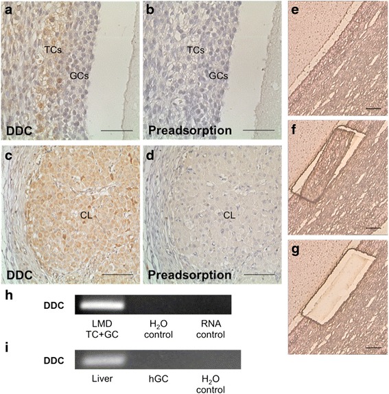
Fig. 4.

DDC in human ovarian tissue. a In human ovarian tissue TCs are positive for DDC in an immunohistochemical staining. b The pre-adsorption control is devoid of staining. c Cells of the human corpus luteum are positive for DDC using immunohistochemistry. d Pre-adsorption abolished staining of the corpus luteum. e–g Micrographs of human ovarian tissue before (e), during (f) and after (g) LMD. TCs and GCs of the follicle wall were excised. After RNA extraction a RT-PCR was performed. Bars indicate 50 μm (a and b) and 100 μm (c–g). h RT-PCR and sequencing showed, that DDC mRNA is present in samples of human TCs/GCs. All controls (input of H2O instead of cDNA and RNA instead of cDNA) were negative. i DDC mRNA is absent in cultured human GCs (hGC; pool of seven preparations). Human liver cDNA was used as positive control. Controls (input of H2O instead of cDNA) were negative
