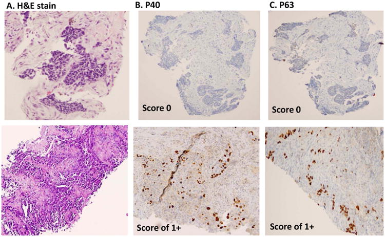
Figure 1.


Semi-quantitative scoring of P40 and P63 in lung SqCC. A, histomorphology of SqCC on H&E slide. B, immunostain of P40, and C, immunostain of P63. The upper panel shows score 0 (negative staining patterns), the mid panel shows score 1+ (focally staining patterns), and the lower panel shows score 2+ pattern (diffusely staining patterns). All photos are taken at 20× magnification.
