Fig.5.
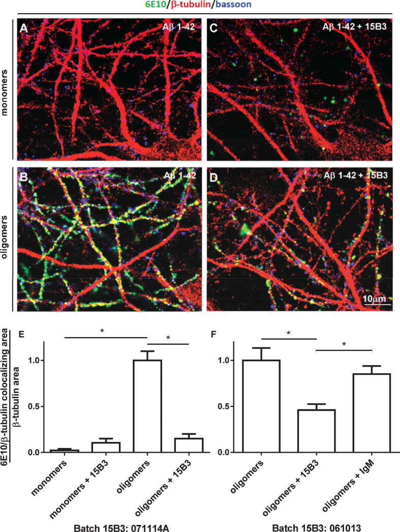
Effect of 15B3 on the binding of Aβ42 oligomers to rat hippocampal neurons—A–D) Representative images obtained exposing 12–15 DIV hippocampal neurons for 1 h to solutions containing (A) Aβ42 monomers or (B) Aβ42 oligomers. The final concentration of Aβ42 was 1 μM in both cases. C, D) Neurons exposed to 1 μM Aβ42 monomers or oligomers pre-incubated for 30 min with 15B3 (batch # 071114A, 10 nM). Neurons were washed, fixed with 4% paraformaldehyde and stained using the following antibodies: mouse anti-Aβ, 6E10 (green), rabbit anti-β tubulin (red) and guinea pig anti-Bassoon (blue). E) Corresponding quantification of 6E10 binding to cultured neurons expressed as colocalizing area between 6E10 and β-tubulin, relative to total β-tubulin. Mean ± SEM of 20 fields from two independent experiments, *p < 0.05 Dunn’s test after Kruskal-Wallis One Way Analysis of Variance on Ranks (this statistical analysis was used because the normality test failed). F) Quantification of a third experiment with 15B3 batch # 061013 and control IgM, both 10 nM. Mean ± SEM of 10 fields, *p < 0.05 Holm-Sidak test after One Way Analysis of Variance (used because the normality test passed).

