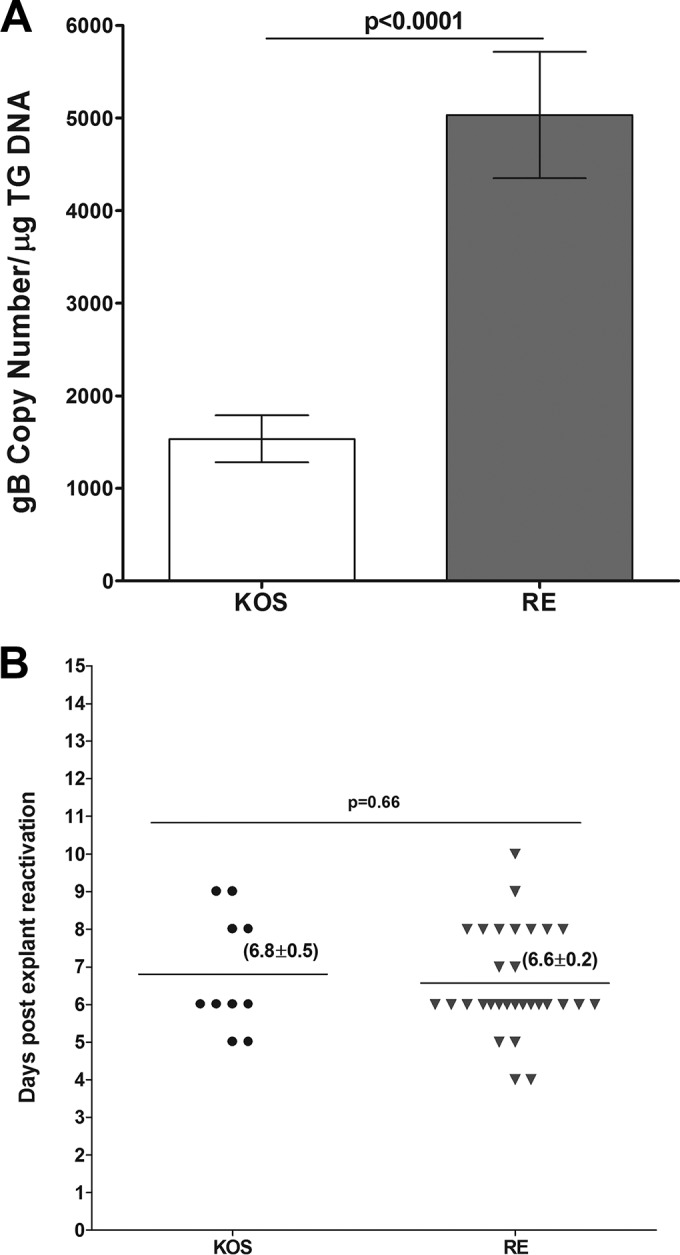
FIG 6.

Level of latency and duration of explant reactivation following ocular infection of mice with avirulent strains of HSV-1. Cornea from wt C57BL/6 mice were scarified before ocular infection and then were infected ocularly with 2 × 10⁵ PFU per eye of HSV-1 strains KOS and RE. On day 28 p.i., TG from infected mice were harvested for qPCR and explant reactivation. (A) gB DNA in latent TG. Quantitative PCR was performed on each mouse TG. GAPDH expression was used to normalize the relative expression of viral (gB) DNA in the TG. Each point represents the means ± SEM from 20 TG. (B) Explant reactivation in latent TG. Each individual TG from infected mice was incubated in 1.5 ml of tissue culture medium at 37°C, and the presence of infectious virus was monitored for 15 days as described for Fig. 2. For each virus, 20 TG from 10 mice were used. In KOS-infected mice, only 50% of latent TG were positive for the appearance of CPE, while in the RE strain 90% of TG showed CPE. Numbers indicate the average time (±SEM) that the TG from each group first showed CPE. (A) gB DNA; (B) explant reactivation.
