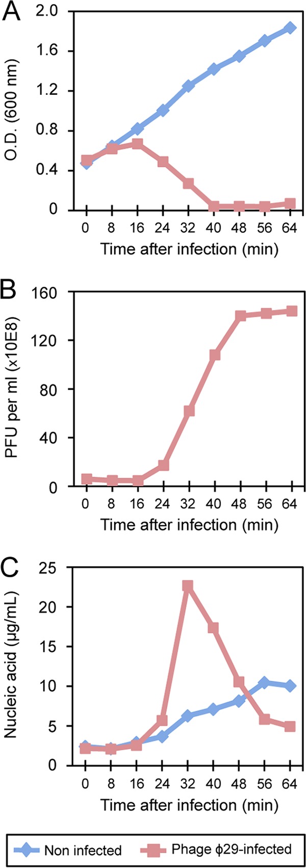
FIG 1.

Analysis of host growth, phage virions, and nucleic acid content throughout the infection cycle of phage ϕ29 in B. subtilis. (A) Phage-mediated lysis of bacterial cultures. Lysis was monitored by measuring the OD600 values for samples taken at the indicated times after infection. (B) The ϕ29 virions were counted as PFU per ml of culture. One hundred-fold-diluted cultures were grown in LB medium containing magnesium at 37°C and then infected at an OD600 of 0.45 with ϕ29 at a multiplicity of 5. (C) The amount of nucleic acid detected at the indicated times postinfection was quantified by using a 2100 Bioanalyzer (Agilent Technologies), and values are expressed as micrograms of nucleic acid per ml of culture. The graphics are representative of four independent experiments.
