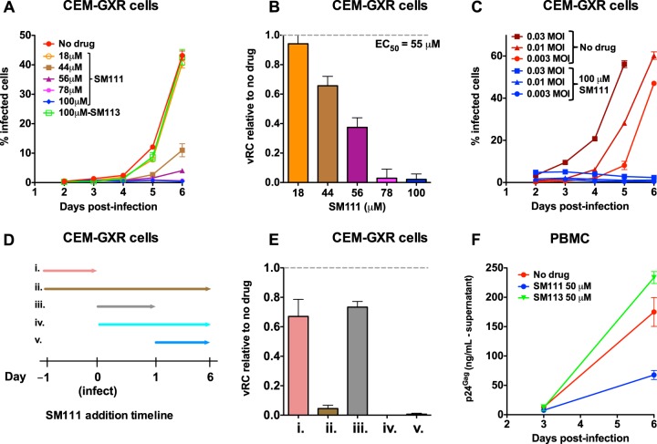
FIG 3.
SM111 inhibits in vitro replication of HIV-1NL4.3. (A) CEM-GXR cells were infected with HIV-1NL4.3 for 24 h (MOI = 0.003), washed, and cultured in the absence or presence of SM111 or SM113 at the indicated concentrations. Cellular GFP, a marker of viral infection, was monitored on days 2 to 6 by flow cytometry. (B) Viral replicative capacity (vRC) of HIV-1NL4.3 in CEM-GXR cells in the presence of indicated SM111 concentrations, normalized to viral spread in the absence of compound. (C) Replication of HIV-1NL4.3 in CEM-GXR cells infected at MOIs of 0.03, 0.01, and 0.003 in the absence or presence of 100 μM SM111. (D) Schematic of experimental plan for results shown in panel E, indicating the times of addition and removal of 100 μM SM111 in CEM-GXR cells relative to infection with HIV-1NL4.3 (MOI = 0.003) at time zero. (E) vRCs of HIV-1NL4.3 in the absence or presence of 100 μM SM111, annotated (i to v) as shown in panel D. (F) PBMC were infected with HIV-1NL4.3 for 24 h (MOI = 0.003), washed, and cultured in the absence or presence of 50 μM SM111 or SM113. Supernatant levels of p24Gag were monitored by ELISA on days 3 and 6 postinfection. For panels A, C, and F, data are representative of three independent experiments performed in triplicate (from three different donors in panel F). For panels B and E, histograms and error bars represent means ± SD calculated from three independent experiments performed in triplicate.

