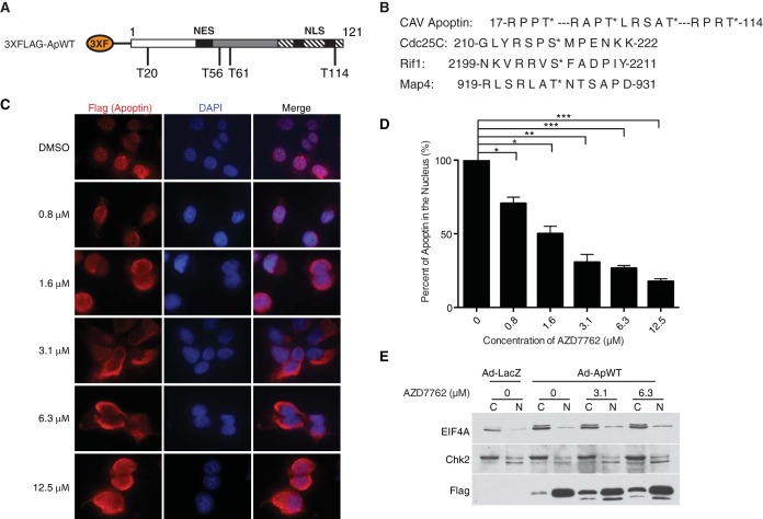
FIG 1.
Treatment with the pan-Chk inhibitor AZD7762 impairs nuclear localization of apoptin in a dose-dependent manner. (A) Schematic of 3×Flag-apoptin showing possible Chk1 and Chk2 phosphorylation sites (not drawn to scale). (B) Comparison of the amino acid sequence of apoptin with those of known checkpoint kinase substrates. Asterisks indicate that the preceding amino acid is the one that is phosphorylated (i.e., the threonine); hyphens indicate a stretch of irrelevant amino acids between the sequences that are shown. (C) H1299 cells were infected with Ad-Apwt and treated with DMSO or the indicated concentrations of AZD7762. At 24 h postinfection, the cells were processed for Flag immunofluorescence. (D) Quantification of cells displaying nuclear apoptin was determined by scoring at least 100 Flag-positive cells per condition. *, P ≤ 0.05; **, P ≤ 0.01; ***, P ≤ 0.0005. The data are presented as means and standard errors of the mean (SEM). (E) Cells were prepared as for panel C but fractionated into nuclear (N) and cytoplasmic (C) fractions and then analyzed by SDS-PAGE and Western blotting. Numbers indicate the concentrations of AZD7762 with which the cells were treated.

